“Sheer depth of analytical possibility” - Using the Phasefocus Livecyte at St George’s University of London
10/08/2021
Why St George's University of London chose the Phasefocus Livecyte
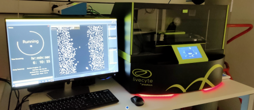
Greg Perry is Image Resource Facility Microscopy Manager at St George’s University of London, where a Phasefocus Livecyte was installed earlier this year. The Livecyte Kinetic Cytometer uses patented Ptychographic Quantitative Phase Imaging (QPI) technology for a range of label-free assays.
The Image Resource Facility
The Image Resource Facility at St George’s is a core facility resource including Light Microscopy, FlowCytometry and Histology. The facility supports a variety of different research including groups looking at:
- Vascular biology - how cells migrate in response to a specific stimulus that has been -identified in the formation of blood vessels.
- Prostate cancer – what possible chemoattractants may be involved in prostate cancer
formation. - Lymphoedema - wound healing assays and scratch assays on lymphatic endothelial cells.
Why the Livecyte
Greg said: “The main reason we chose the Livecyte was the combination of technology and custom-built analysis. The system is designed around assays, which takes away the requirements from users to delve into the technical setup. We chose it to save time not just in set up but analysis too.”
Removing barriers to entry
“Another reason is that the Livecyte removes barriers to entry for junior students. The time scale of undergraduate projects is often quite limited, and complex training of techniques can take weeks. Students may not get to grips with particular microscopy techniques within that timescale, but the Livecyte removes that delay and enables students to focus on their studies and contribute to a bigger picture of research. The process of using the Livecyte has been exceptionally straightforward and it is incredibly simple to get to grips with. More often than not, there is no guidance needed when a new user comes to use the Livecyte because the system is so intuitive in the setup."
“Someone who’s never seen the Livecyte before can get 90% of the assay ready to go before needing any additional guidance. Students can get to work within 20 minutes of placing the assay; it makes work much more reliable for me and the principal investigator who manages the students.”
What the users say
Greg said: “One professor has been using the Livecyte on a daily basis and his workflow has become so streamlined that he has managed to get a few months’ worth of work done in just one. Typically, his analysis and manual tracking would be very time consuming, but the Livecyte does this automatically – revolutionising his work. Some projects have been conceptualised but not started due to lack of personnel, however, the Livecyte now allows them to revisit those projects and seek funding to run them.”
“Sheer depth of analytical possibility”
Greg added: “I think the sheer depth of analytical possibility is really starting to dawn on people. You can generate incredibly complex outputs but you've got the surface level analysis too. It ticks all boxes! The quality of the simple, super easy to grasp the operation of the system, but the sheer depth of what you can do with it, is very inspiring.”
LabLogic and Phasefocus partnership
LabLogic is Phasefocus’ UK partner and Lucy Farmer is Life Sciences Product Specialist, looking after the cell imaging range.
Greg said: “The service from LabLogic and Phasefocus has been excellent and incredibly responsive.
“Lucy synchronized the whole process very well – between the sales and logistics side from LabLogic and the technical side from Phasefocus.”
“Overall, I’m really pleased with my experience so far.”