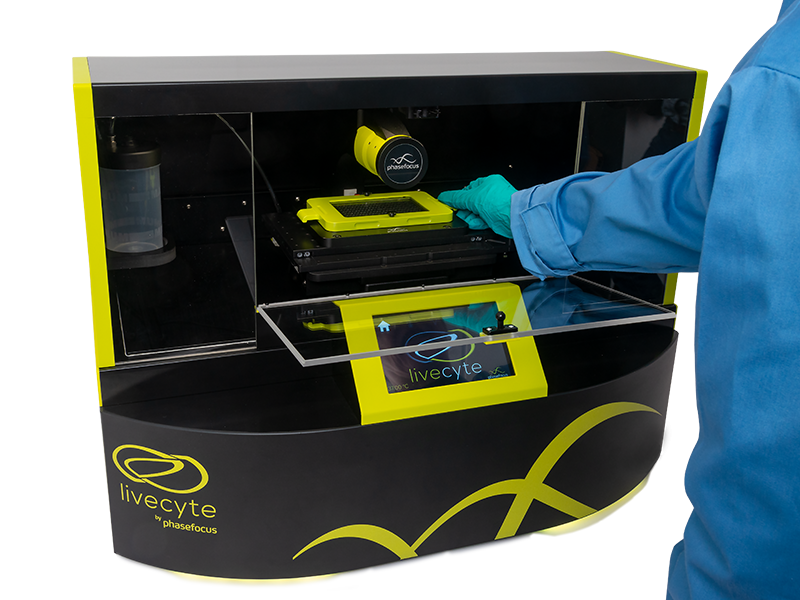
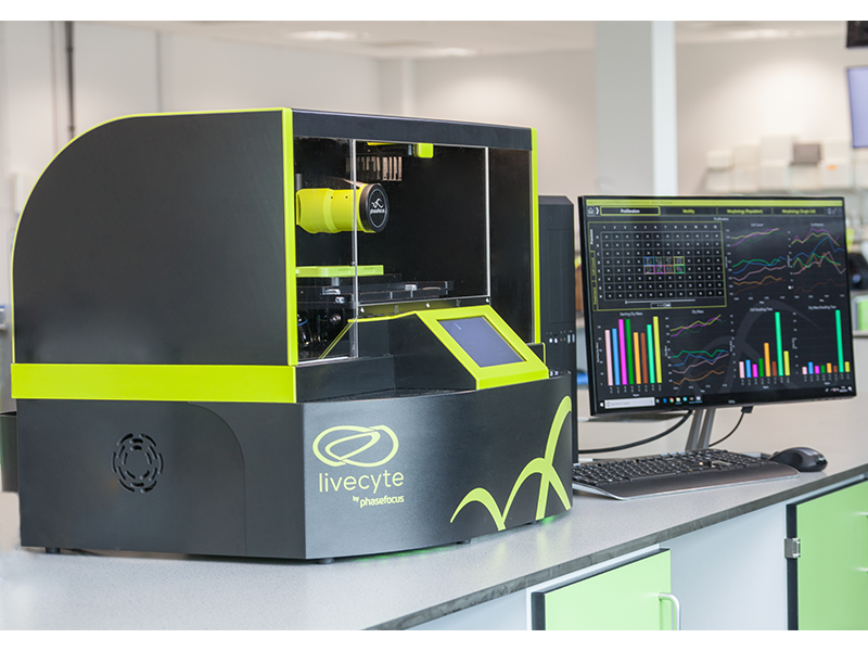
The Livecyte kinetic cytometer produces exceptionally high contrast time-lapse videos using Phasefocus’s patented Ptychographic Quantitative Phase Imaging (QPI) technology for a range of label-free assays with or without up to seven channels of complementary fluorescence.
Automated single-cell tracking of even the most sensitive cells quickly reveals subtle phenotypic differences in unperturbed cell populations.
Easy-to-use dashboards present coherent and concise results from up to 96 wells at a time whilst retaining the ability to investigate individual cell behaviour and outlying characteristics.
Livecyte Kinetic Cytometer Applications
Explore the tabs below to learn more about the various Livecyte applications including videos, application notes and more.
Cell Growth and Proliferation
Livecyte measures individual cell growth independently from proliferation
Cell growth is more complex than just cells multiplying
They can increase in size without dividing, they can divide asymmetrically, they can grow to a certain size then stop. All these aspects of cell growth are lost with most live cell assays. Not with Livecyte
Wound Healing
Directly measure cell motility with individual cells
Separate out migration from proliferation in a single experiment
Observe differences between leading edge and following cells
Correlate motility to morphological differences
Read the Application Note 'Single Cell Scratch Assay' from Phasefocus here.
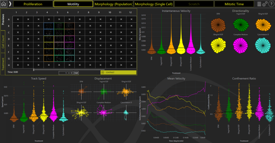
Cell Motility
Livecyte automatically tracks cells, even in a 96-well plate
Just seed your cells, monitor with Livecyte and a full range of cell migration parameters will be generated
Compare the speed, directionality, confinement and heterogeneity of different populations
Oncology
Directly measure mitotic time with individual cells
Measure random and chemotactic migration
Identify and characterise differences in subpopulations in heterogenous primary cultures
Correlate motility to morphological differences
Generate statistically valid results from multiple repeats in 96-wells
High-resolution large fields-of-view with no stitching
Perform assays on Matrigel and standard plates
Automated post-acquisition focus ensures consistent image quality for every well
Simple, yet powerful analysis of tube formation metrics with Application Dashboard outputs
Stem Cells
Identify morphological phenotypes
Watch and quantify cell differentiation
Monitor stem cell populations, for many days, without perturbing cell behaviour
Image through plastic and coated plates
Image on extracellular matrices
Cells are still viable after imaging
Livecyte Kinetic Cytometer Videos
Livecyte: Multiple Assays In Every Experiment
See how Livecyte maximises every experiment, saving time and conserving precious cells.
This short 80-second video illustrates how proliferation, morphology and motility metrics are automatically generated and compared, down to single-cell level, in every Livecyte assay.
Livecyte: Fluorescence Gating
Want to analyse mixed cell populations and co-cultures?
This short 90-second video shows how Livecyte combines both label-free and fluorescence imaging to easily identify distinct sub-populations of cells and compare behaviour within mixed populations, by gating on fluorescence labels.
Livecyte: Explore new cell relationships
Go from plate, to population, to individual cell using the "Explore" functionality, where intuitive visual data representations will let you interrogate your cells, gate sub-populations and investigate the links between data-points and images.
Livecyte: Long Term - Large field of view (FOV)
With Livecyte, track your cells for periods of days or weeks, without the need for image stitching to track every cell through its lifecycle.
Existing Phasefocus Livecyte users
Click the tabs below to learn more about our customers and their applications for the Livecyte.
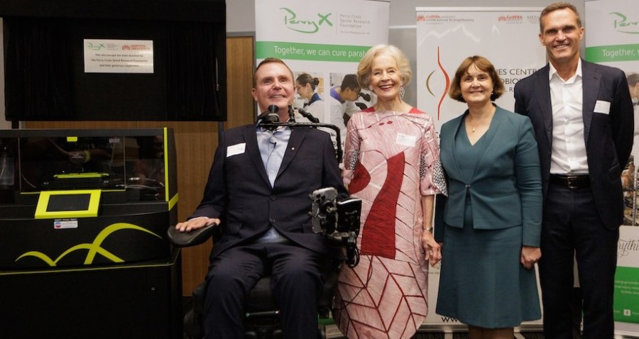
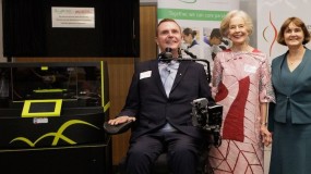
Clem Jones Centre, Australia
Researching therapies for spinal cord injuries
The Clem Jones Centre for Neurobiology and Stem Cell Research at Griffith University in Brisbane is using the Livecyte to research therapies for spinal cord injuries. Professor James St John heads the Spinal Injury Project based at the centre with research funded by the Perry Cross Foundation. The project is looking to begin human clinical trials to test cell transplantation therapy and repair spinal cord injury, ultimately leading to restoration of function. The Livecyte will be used to perform critical cell analysis and develop a pre-screening process before cell transplantation and human clinical trials.
You can learn more about this ground-breaking research and the role of the Livecyte in it by clicking the button below.
Representative image of the Livecyte's Cell Motility Dashboard. Image analysis, cell segmentation and tracking data across multiple wells are automatically curated into tangible cell migration metrics.
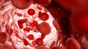
The Livecyte is being used by the University of Nottingham at its Centre of Membrane Proteins and Receptors (COMPARE) to noninvasively monitor the morphological phenotype and behaviour of immortalised endothelial cells. As fluorescent labels can perturb the natural behaviour of cells, the Livecyte is an excellent instrument for cell research because it uses ptychographic quantitative phase imaging (QPI) for label-free assays.
Assistant Professor at the School of Pharmacy Dr. Laura Kilpatrick is investigating if endothelial cells behave similarly to human umbilical vein endothelial cells (HUVEC), and that genetic mutation was not altering their phenotype. A hallmark of the endothelial phenotype is their ability to form new blood vessels in a process called angiogenesis.
Click the button below to find out what Dr. Kilpatrick had to say about the Livecyte and her research.
The Livecyte is being used at the University of York’s core imaging facility to answer a variety of scientific questions previously unanswered by modern imaging techniques. In our most recent video, we spoke to several researchers from the university’s Imaging & Cytometry Laboratory to find out how the Livecyte has enabled their research.
Dr. Will Brackenbury carries out research in cellular electrophysiology and has been using the Livecyte to generate high quality, reproducible data. His work primarily focuses on understanding ion movement through channels in cell membranes, with the hope to identify these channels as therapeutic targets in the future.
Dr. Laura Wiggins uses the Livecyte to study cell phenotyping; the ability to profile a cell in terms of its shape, size, texture, and motility. Automated single cell tracking of even the most sensitive cells quickly reveals subtle phenotypic differences in unperturbed cell populations.
Watch our video above or click the button below to find out more.
Identifying the role of HECTD2 in the cellular proliferation of melanoma cells
Dr. Eleonora Ottina and her team at the Francis Crick Institute have used the Livecyte to identify a new role of the ubiquitin ligase HECTD2 in the cellular proliferation of melanoma cells, which has been suggested as controlling inflammation and immunity in melanoma patients. Ottina et al. studied the gene-expression profile of melanoma patients and discovered several cell cycle genes associated with HECTD2 expression, which were upregulated.
You can find out about the team's research by clicking button below.
TheLivecyte has been installed at Texas Tech University’s core imaging facility at the Amarillo Research Building. Dr. Constantinos Mikelis, Adjunct Associate Professor, and Dr. Ulrich Bickel, Professor in the Department of Pharmaceutical Sciences, have been using the cytometer for cancer and vascular research in pharmaceutical sciences.
Click the button below to watch an interview with Dr. Mikelis and Dr. Bickel about their work.
High-content imaging and cell analysis in the study of melanocytes
The Phasefocus has been used by Dr. Robert Judson-Torres’s research group at the Huntsman Cancer Institute in Utah. Working in collaboration with McNeal et al. from the University of California, they investigated how melanocyte cells switch from stable nevus cells to cancerous melanoma cells, and how previously benign melanomas may re-exhibit growth after cell cycle arrest.
Click the button below to read more about this collaborative research.
Greg Perry is Image Resource Facility Microscopy Manager at St George’s University of London where a Phasefocus was installed to support a research groups looking at vascular biology, prostate cancer, and lymphoedema.
Click the button below to watch our interview with Greg.
The new assay addresses challenges faced by researchers in investigating the efficacy and cytotoxicity of engineered T-cells in killing target cancers…