Macrophage immunology assays using the Phasefocus Livecyte
01/08/2022
Quantifying phagocyte behaviour down to the single-cell level
The Livecyte kinetic cytometer by Phasefocus combines high contrast label-free imaging with correlative fluorescence and automatic cell tracking for quantifying phagocyte behaviour down to the single-cell level.
This enables users to:
- Automatically quantify macrophage phenotypic behaviour label-free to measure immune response
- Quantify to the single-cell level, giving a more accurate measure of phagocytosis than standard population-level analyses
- Increase the accuracy to reveal subtle behavior changes from how phagocyte activity changes over time to saturation
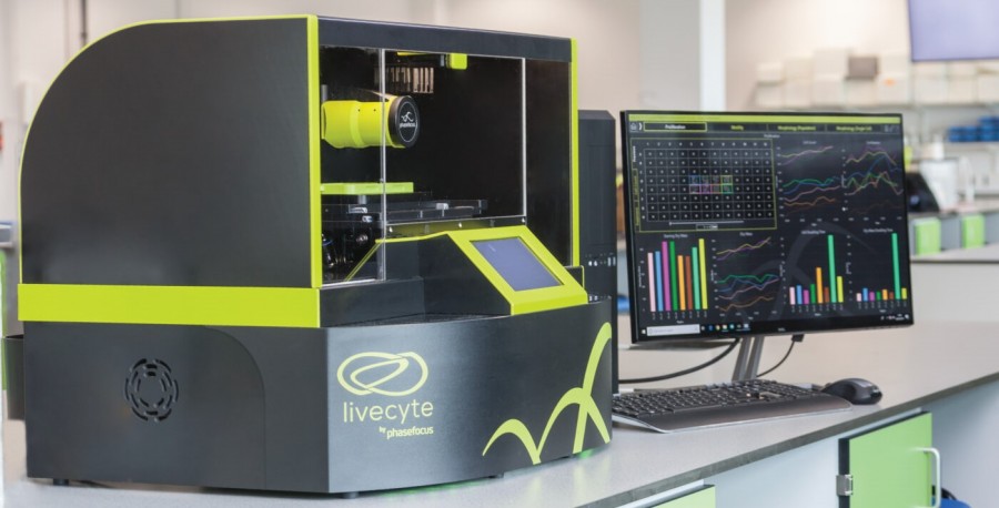
The Livecyte kinetic cytometer by Phasefocus
Quantifying phenotypic changes to measure immune response
Macrophages are known to change their behaviour or state in response to specific functional initiatives. Mediators interferon-γ (IFNγ) and bacterial lipopolysaccharide (LPS) promote a more active pro-inflammatory M1 phenotype, but measuring this response is challenging.
The quantitative phase images produced by Livecyte are ideal for single-cell segmentation and tracking that enables automatic quantification of phenotypic behaviour such as cell morphology, motility, and proliferation. This allows macrophage immune response to be measured in an assay-relevant format by the quantified change in phenotypic behaviour.
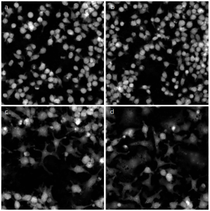
Quantitative phase images
Quantitative phase images of a) untreated macrophages and those treated with b) IFNγ (10ng/ml), c) LPS (10ng/ml) and d) a combination of both.
Images were taken at x20 magnification at 40hrs and illustrate the cellular and sub-cellular morphological changes observed.
You can read more about quantifying macrophage activation and proliferation by clicking here.
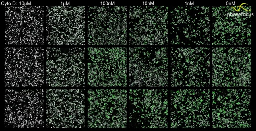
Single-cell phagocytosis measurement
A key function in macrophage immune response is the phagocytosis of foreign bodies or pathogens. Traditionally this is quantified by measuring the fluorescence expression of an appropriate biomarker across a whole image. However, this population-level approach oversimplifies complex single-cell level mechanics and population heterogeneity.
Livecyte quantifies correlative fluorescence expression down to the single-cell level to eliminate confounding effects from cell proliferation and preserve the heterogeneity of the response. On Livecyte, fluorescence imaging can be performed intermittently as individual cells are tracked using the Quantitative Phase Imaging (QPI) modality, substantially reducing phototoxicity for the assay.
You can learn more about the phagocytosis measurement of bioparticles by clicking here.
Reveal how phagocyte activity changes over time
Efferocytosis, the phagocytosis of apoptotic cells, is critical for the maintenance of tissue health and reducing inflammation. Livecyte can combine the quantification of phagocytosis and the measurement of macrophage phenotypic behaviour in a single experiment, which can lead to insights not possible from either assay in isolation.
For example, in AN019 we correlate phagocyte activity with changes in macrophage motile behaviour and label-free growth and proliferation to identify changes in individual phagocyte activity over time and potential phagocyte saturation.
You can learn more about the phagocytosis of apoptotic cells by clicking here.
Find out more
You can learn more about the Livecyte by clicking the button below to request a guided demonstration with a product specialist.