(Application Note) Phenotypic Profiling of Fission Yeast: A Model Organism
14/06/2021
(Application Note) Phenotypic Profiling of Fission Yeast: A Model Organism
Yeast is a commonly used model organism for studying underlying cellular processes, in particular cell morphology, division regulation and cell-cycle progression. Researchers want to quantify these behaviours and how they change over time, as a function of treatment or between different genetic strains.
Quantifying these changes would normally require fluorescent labeling, introducing phototoxicity effects, or manual analysis from brightfield or phase contrast imaging modalities, which lack the necessary contrast for automated analysis.
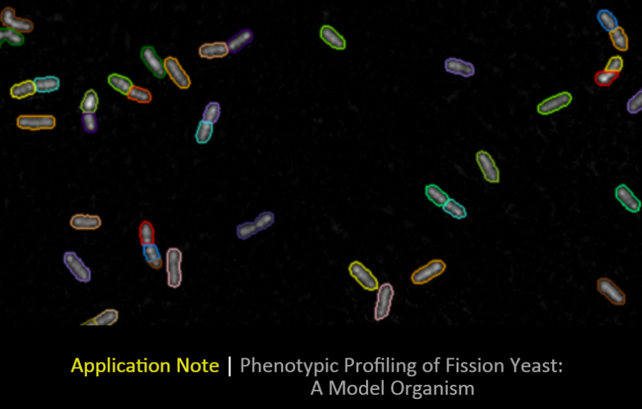
Why Livecyte?
Livecyte's higher NA objective lenses enable it to utilise Ptychography, a quantitative phase imaging (QPI) technique, to produce high-contrast images without the need for fluorescent labels even on smaller biological organisms such as yeast. The enhanced contrast enables the automatic segmentation and tracking of individual cells, as well as a quantitative measure of cell morphology.
Additionally Livecyte can measure changes in cellular dry mass. This provides a measure of cell growth allowing researchers to independently separate out effects on cell-cycle progression from cell growth.
In the app note...
- Investigate morphological changes in different yeast strains.
- High-contrast label-free imaging for robust single cell segmentation.
- Extract a wealth of single cell and population level metrics including Dry Mass, Sphericity, and length:width ratio.
Download the app note highlighting quantification of yeast morphological differences due to genetic mutation here.