Determining cell proliferation after being exposed to GPC1 with the Absorbance 96
29/01/2024
The Byonoy plate reader has been used in a recent breakthrough study by Cheng et al. to investigate glypicans in tumorigenesis
To find new biomarkers, prognostic factors, and therapeutic strategies for cancer, it is essential to decipher the mechanisms involved in communication between cancer cells and surrounding tissues. GPCs (Glypicans) are anchored proteins located on the cells surface. Due to their cell surface localisation and specific structures, GPCs can interact with a wide range of proteins, including morphogens, growth factors, cytokines, chemokines, ECM proteins, and adhesion molecules. Such interactions have been shown to play key roles in neoplastic growth and neovascularisation.
Understanding the role of GPC1 in neoplastic behaviour
Studies show that GPC1 plays a role in neoplastic behaviour by modifying mitogenic signalling pathways exerted by different growth factors. Furthermore, GPC1 has been shown to undergo recycling and contribute to clearance of oxidative damaged proteins in cancer and neuronal cells. With signal pathways modified and oxidised proteins being cleared out of cancer cells, it is clear that the presence of GPC1 causes cancer cell proliferation by inhibiting normal apoptosis cycles.
Investigating the differences in gene expression patterns of the GPC family
One study conducted by Cheng et al. investigated the role of GPC1 in tumorigenesis. Using data available from The Cancer Genome Atlas (TCGA), the study systematically investigated differences in gene expression patterns of the GPC family in normal and malignant tissues. Employing a series of genetic perturbation experiments including knock-out, knock-down and overexpression assays, the study discovered that downregulation of GPC1 results in attenuation of cell proliferation across different in vitro cancer models.
Measuring cell density with the Absorbance 96
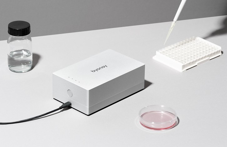
The Byonoy Absorbance 96 was used by Cheng et al. to determine cell proliferation after being exposed to GPC1. Cells were seeded in 96-well microculture plates and after 24 hours, the cells were either left untreated or were treated with CRISPRcontrol, CRISPR/Cas9GPC1, siRNAmock, siRNAGPC1, GFP-GPC1 or anti-GPC1 polyclonal antibody. After 3-4 days, the cell density was measured by staining of the nuclei with 0.1% (v/v) crystal violet, followed by measurement of the amount of bound dye at A595 nm using the Byonoy microplate reader. Cell proliferation rate was calculated using Byonoy Absorbance 96 software alongside MS Excel and the relative cell number was calculated as % of untreated cells or controls (CRISPRcontrol or siRNAmock).
Find out more
You can learn more about the Absorbance 96 by clicking the button below to speak to a product specialist directly to request a guided demonstration or click here to read the full study.