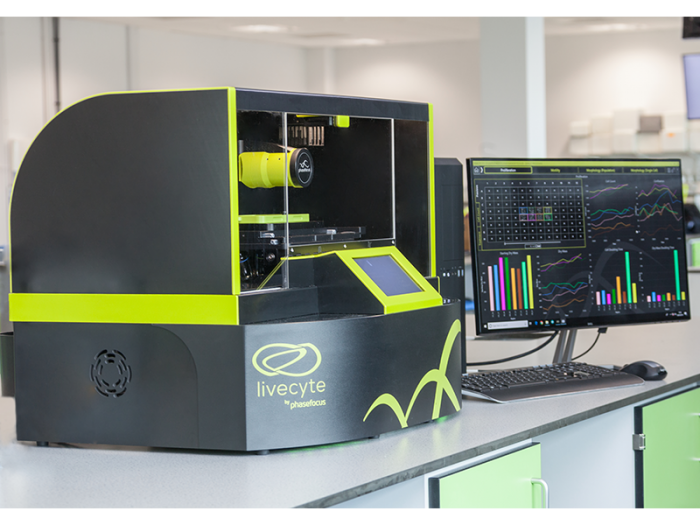
Phasefocus Livecyte Kinetic Cytometer at the Huntsman Cancer Institute
18/01/2022
Using Livecyte’s high-content imaging and cell analysis in the study of melanocytes
The Phasefocus Livecyte Kinetic Cytometer has been used by Dr. Robert Judson-Torres’s research group at the Huntsman Cancer Institute in Utah. Working in collaboration with McNeal et al. from the University of California, they investigated how melanocyte cells switch from stable nevus cells to cancerous melanoma cells, and how previously benign melanomas may re-exhibit growth after cell cycle arrest.
The Huntsman Cancer Institute had very specific requirements that conventional imaging could not fulfill, but Livecyte could by measuring individual cell growth independently from proliferation. This helped them to determine at what point the cell cycle arrest was occurring.
Ptychographic Quantitative Phase Imaging
McNeal et al. wanted to characterise the growth and proliferation of the melanocyte cells with tumorigenic potential and drug resistance. Conventional imaging could not capture this in-depth because it relies on cell counts and confluence. This obscures abnormal growth patterns, such as accumulation of dry mass where cells are getting larger, but not proliferating at the same rate.
Because Livecyte uses patented Ptychographic Quantitative Phase Imaging (QPI), it allows the collection of high-contrast, label-free images where pixel intensity is directly proportional to cell dry mass. This enabled robust cell segmentation and tracking that lead to accurate single-cell measurements of proliferation.
Fluorescent imaging
As well as QPI, Livecyte also allows low-intensity fluorescent imaging that can be taken intermittently and correlated to the phase image. This reduced the levels of phototoxicity that commonly occurs during fluorescent imaging and enabled the cells to grow with minimal disturbance.
Reliable results with limited sample sizes
Cultivating the sample cells was very difficult because they had undergone cell cycle arrest. They had not been growing or proliferating so each specimen only provided one or two 96-well plate experiments each. However, because Livecyte can segment every cell and uses each individual one as a data point, it was possible for the team to extrapolate enough reliable and significant cell proliferation data for study. This would not have been possible if each well had been treated as a single data point, which is what high throughput analyzers do.
Find out more
By using Livecyte, McNeal et al. were able to identify both proliferation arrest in their melanocytes and the continuation of cell growth, implying a cell cycle block, as well as a mutation that proves key in the conversion of stable nevi into melanoma.
You can learn more about the science of the study at the Huntsman Cancer Institute by visiting the Phasefocus website or you can learn more about the Livecyte by clicking the button below to speak with our product specialist directly and have a one-to-one guided session.